Essentials Of Clinical Anatomy Of The Equine Locomotor...
| Item Information | |
|---|---|
| Item#: | 9781498754415 |
| Author | Denoix, Jean-Marie |
| Cover | Hardback |
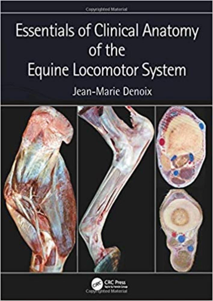
Essentials ofClinical Anatomy of the Equine Locomotor System presents a unique photographic record of dissections showing the topographical anatomy of the locomotor system of the horse. Readers of this book will be able to see the position and relationships of the bones, joints, muscles, nerves and blood vessels that make up each region of the forelimb, vertebral column and hindlimb.
Key features:
Important features of regional and topographical anatomy are presented using full-color photos of detailed dissectionsAnatomy is presented in a clinical contextPreparations of cross-sectional anatomy facilitate interpretation of diagnostic imaging, such as ultrasonography, MRI images and CT scansAll dissections are of fresh material, rather than preserved specimens, to demonstrate the appearance of tissues in the living animal, or at post mortem autopsyThis new atlas is essential for anybody involved in detailed anatomical study, complex lameness evaluation or advanced imaging techniques in horses. It will be a useful guide for veterinary students, and a reference for equine vets in practice.
Preface. Chapter 1. FORELIMB: Foot, Pastern, Fetlock, Metacarpus, Carpus, Forearm, Elbow, Arm, Shoulder. Chapter 2. VERTEBRAL COLUMN: Neck, Back, Pelvis. Chapter 3. HINDLIMB: Digital area, Fetlock, Metatarsus, Tarsus, Crus, Stifle, Thigh, Hip. Index.